


In vitro evaluation of dentin marginal adaptation of three root-end filling materials inserted with and without surgical microscope
Bernardo Mattos Almeida, Ernani da Costa Abad, Hélio Rodrigues Sampaio Filho, Juliana de Oliveira Zóffoli
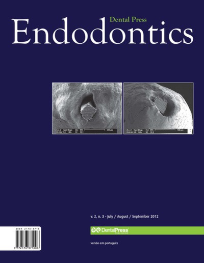
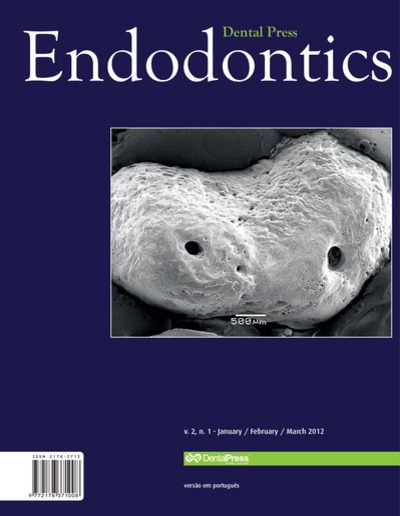
Objective: Periradicular surgery is the procedure of choice when one aims to solve problems or complications which conventional endodontic therapeutics has not been able to solve. Surgical therapy comprises a number of procedures, among which retrofilling. The aim of this study was evaluating the degree of dentin marginal adaptation of root-end filling materials, as well as ascertaining the effectiveness of optical microscopic usage in the insertion of these materials. Methods: Sixty upper canine teeth were selected, apicectomized and then rood-end cavities were prepared with the help of ultrasonic tips. The specimens were divided according to the material used: White MTA Angelus®, Super-EBA® and Sealapex® + White MTA Angelus®, it being that optical microscope was used in half of the samples of each group. All samples were processed and evaluated by scanning electronic microscopy (SEM). Results: The three materials tested presented satisfactory marginal adaptation. The use, or not, of the optical microscope, did not change the degree of adaptation of root-end filling materials evaluated in the present study. Conclusion: All materials tested (White MTA Angelus®, Super-EBA®, and Sealapex® cement added to White MTA Angelus®) proved efficient regarding the issue evaluated, dentin marginal adaptation. The use of the optical facilitated insertion of root-end filling materials, due to better illumination and magnification. However, it did not promote any difference in the materials marginal adaptation to root-end cavities, when compared with its non-utilization.
Keywords: Retrograde obturation. Scanning electron microscopy (SEM). Endodontics. Oral surgery.
How to cite: Almeida BM, Abad EC, Sampaio Filho HR, Zóffoli JO. In vitro evaluation of dentin marginal adaptation of three root-end filling materials inserted with and without surgical microscope. Dental Press Endod. 2012 Oct- Dec;2(4):50-5.
Friday, April 19, 2024 06:50