


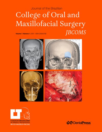
Surgical treatment of sialolith on submandibular gland duct
PAULO MATHEUS HONDA TAVARES, LETÍCIA DOS SANTOS NASCIMENTO, VALBER BARBOSA MARTINS, MARCELO VINICIUS OLIVEIRA, GUSTAVO CAVALCANTI DE ALBUQUERQUE, SAULO LÔBO CHATEAUBRIAND DO NASCIMENTO, RAFAEL SARAIVA TORRES
Sialoliths are calcified structures that develop in the salivary glands or in their ducts, causing its obstruction and producing inflammation or infection of region. Diagnostic methods include inspection, palpation, radiographic examination, computed tomography and a detailed anamnesis. A 31-year-old male with no systemic alterations presented with complaints of in- creased volume and discomfort in the oral floor during meals. On clinical examination, a volume increase was observed in the right-side buccal floor, and on palpa- tion, a nodule of hardened and mobile consistency was observed. A cone beam computed tomography scan was performed, in which the presence of a hyperdense and cylindrical mass, suggestive of salivary calculus in the region corresponding to the duct of the submandib- ular gland on the right side, was evidenced. The pro- posed treatment was surgical removal, through intra- oral access under local anesthesia and installation of a plastic device to avoid the collapse of the duct. In the postoperative period, the patient had no complaints of pain or discomfort, and the physiological functions were preserved. It was concluded that a quality image examination and evaluation by the dental surgeon are fundamental for definition of the diagnosis and plan- ning of the surgical procedure to be performed.
Keywords: Submandibular gland. Salivary gland calculi. Salivary duct calculi.
How to cite: Tavares PMH, Nascimento LS, Martins VB, Oliveira MV, Albuquerque GC, Nascimento SLC, Torres RS. Surgical treatment of sialolith on submandibular gland duct. J Braz Coll Oral Maxillofac Surg. 2021 May-Aug;7(2):56-62.
Tuesday, May 14, 2024 07:41